
Whitepapers
The latest capabilities of the Diagnostic OS
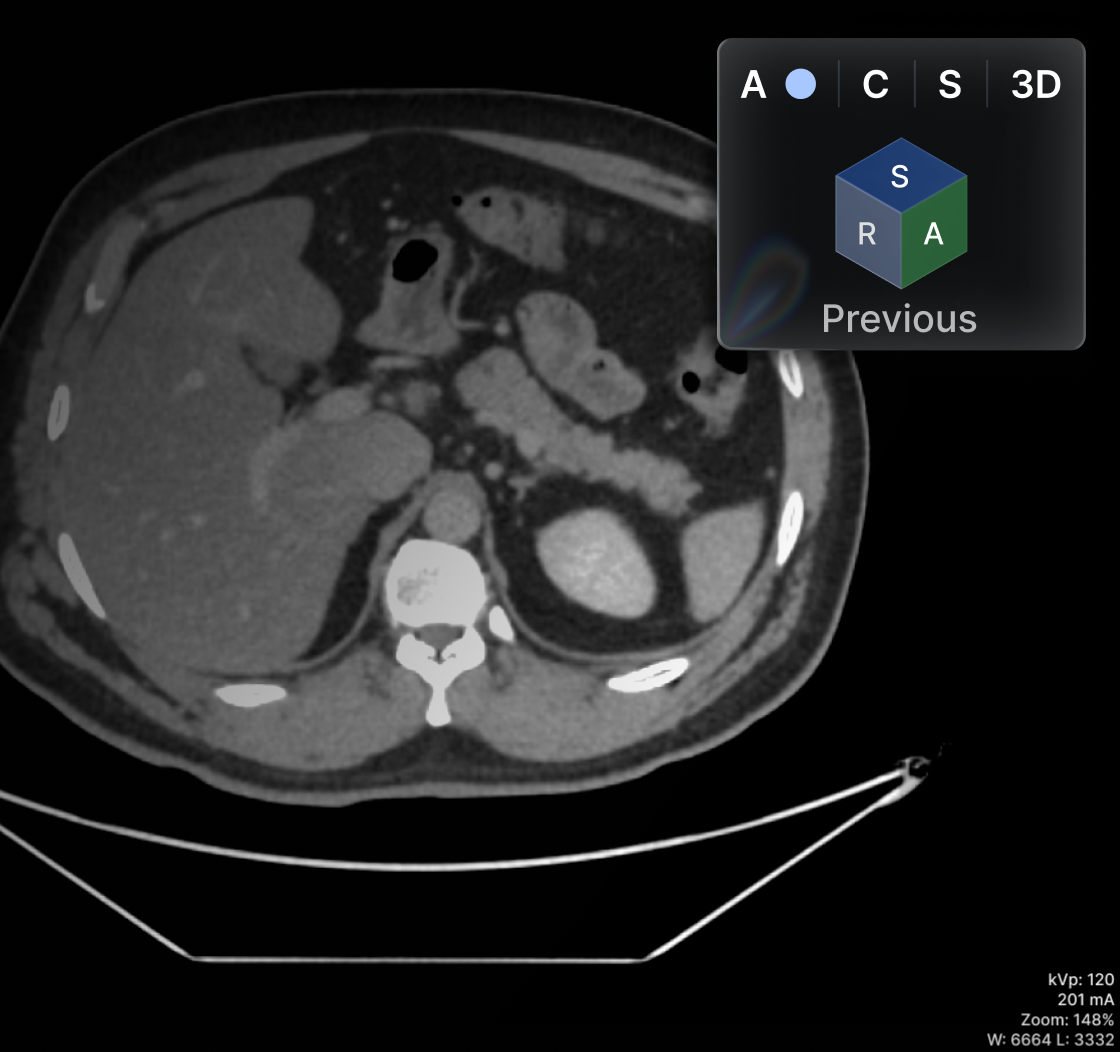
Oblique MPR in the main viewer (not only on the MPR viewer)

.png)
Browse all

Managing a Radiology Laboratory
Increasingly, physicians from different specialties use radiological studies to make more accurate diagnoses for their patients. As a result, these laboratories are in high demand worldwide, with significant growth since the COVID-19 pandemic. Analyzing the body's structure is the best tool for health experts to assess the condition of organs, soft tissue, nerves and much more.
However, for these studies to be of high quality, proper management of the radiology laboratory is essential, and this involves more than just having the best equipment or technical staff. The reason is that, beyond image capture, the management of order, planning, control and direction of all the elements that are part of the healthcare facility also plays a role.
To achieve good management, the first step is to define the laboratory's mission, vision and values. Based on this, administrators, who are not only technicians or radiologists, can develop a work plan to maintain order in the facility, schedule patient appointments, manage records, control invoices, archive studies and design strategies focused on business growth.
Hospital Administration in Radiology
Everything discussed above is closely related to the healthcare specialty known as "hospital administration," which in radiology refers to the complete management of these services. In other words, it refers to optimally managing the waiting times of patients and physicians to obtain imaging results.
It also involves managing patients' historical records, the confidentiality of information, and the complete safekeeping of studies. This is undoubtedly a fairly difficult task when done in the traditional way, that is, with physical X-ray films, which tend to deteriorate over time, are subject to greater human error in the filing process, and require more time to search for as they age.
In this regard, PACS (Picture Archiving and Communication System) systems are an effective technological solution for hospital administration in radiology, since they are digital platforms that make it possible to store, communicate and safeguard imaging studies with the cybersecurity required by industry regulations.
Efficient Radiology Service
By this point, you will notice that a good healthcare facility offering radiology services requires sound management to provide an excellent experience for patients. The key points are:
- Reduced waiting times. Every minute is valuable for patients and healthcare professionals, so image capture and processing must be optimal. This can also increase the facility's productivity.
- Study sharing. Patients also want to have their radiology images on hand to keep their own records, or alternatively to be able to consult with other trusted specialists. Therefore, being able to send these results to patients represents modernization and translates into good service.
- Information security. No one wants their studies disclosed without authorization. For this reason, the systems used in a radiology laboratory must guarantee their confidentiality.
- Backup of clinical records. Although people usually bring their previous studies when they see a doctor, it is also reassuring for them to know that the hospital keeps their records. However, this search must be fast: using only an identification code, whoever handles this task must be able to find the corresponding images and their respective report.
Improvement Plan for Radiology Laboratories
One of the improvement plans for radiology laboratories to enhance their results is through technology. This involves both the use of cutting-edge radiology equipment with excellent resolution for different modalities, as well as systems that streamline storage and communication management activities. PACS fully addresses this last point, since it integrates with the patient database and imaging studies.
In addition, venturing into the digitalization of hospital and radiology services means significant space savings in healthcare facilities, since physical files are no longer needed to store film. Servers, in turn, are the new brain of information. Eva PACS has everything you need to enhance your radiology laboratory.

Patience and Patients: How Radical Empathy Will Transform Healthcare Design
Empathy is a fundamental human quality that refers to the ability to understand and share the feelings of others. Lately, in the world of healthcare, empathy has taken on essential importance in making a difference in the patient experience.
Why can empathy elevate care and benefit institutions as well? Because by ensuring that patients receive enough care to recover from illness or injury, we meet the objective, but only up to that point. Having a basic understanding of patients' feelings still is not enough. To truly transform healthcare, we must raise the standard of quality with radical empathy.
What Does Radical Empathy Consist Of?
Radical empathy is about putting yourself in patients' shoes and seeing the world through their eyes. It is a deep and genuine way of understanding the person, with compassion. This approach to healthcare means authentically understanding their fears, concerns and challenges, in order to offer more personalized and effective care. In this way, by implementing radical empathy, we can achieve truly significant improvements in healthcare.
On the other hand, radical empathy can also help transform healthcare into a more human and personalized experience. By placing the patient at the center of everything, meeting their individual needs becomes a priority throughout the entire process. This compels us, as service providers, to think about and design more effective solutions for the patient.
One example of these solutions would be the use of cutting-edge technologies to improve communication and collaboration between patients and their healthcare providers, doctors, and clinic and laboratory staff at every point of interaction. Another example would be home care solutions that offer a more comfortable and effective service and recovery alternative by sparing patients all the inconvenience of traveling to the healthcare facility.
In addition, applying radical empathy when serving the patient helps reduce the stress and anxiety often experienced when undergoing studies, tests or any other medical intervention. Healthcare can be a stressful and confusing experience, especially for those with no prior experience or knowledge about their condition or treatment. Approaching the patient with radical empathy can help create a more welcoming and understanding environment, minimizing their fears and improving their overall experience.
Patient-Centered Healthcare Design: The Power of Radical Empathy
Radical empathy is a key and valuable tool for transforming healthcare design from now on. By placing patients at the center of everything and authentically understanding their needs and challenges, we are better equipped to design solutions and processes that are more effective, warmer, and more human, in order to deliver exceptional medical care.

History of Radiology: How the Diagnostic Tool Came to Be
Radiology is the branch of medicine concerned with the detection and monitoring of diseases through the use of imaging techniques. Radiologists are medical professionals who specialize in diagnosis and the management of imaging-related patient care. The first use of X-rays dates back to 1896, when German physicist Wilhelm Röntgen discovered them while experimenting with discharge tubes. With his new discovery, Röntgen was able to demonstrate that exposure to these rays produced an image on a photographic plate on the other side of the room where a device known as a vacuum tube was operating. That same year he published his findings on this new type of ray; however, people were slow to recognize the potential medical applications of radiography.
1900s: From Experimentation to Medical Use
The first medical use of X-rays began in 1900, when British physician John T. Scanlon used them to examine the chest and abdomen. In 1901, William Roentgen (Röntgen's surname was shortened to Roentgen when he was admitted to the British Institution of Electrical Engineers) performed the first X-ray examination of the human body. Roentgen's discovery was initially used to examine bones and the skeletal system, but it soon became clear that this new imaging technique could reveal diseases in other organs and tissues, such as the lungs, the abdominal region and the heart. For example, these rays could be used to determine whether a patient had tuberculosis or broken bones. The first hospitals with X-ray departments opened in the United Kingdom, Germany, the United States and France in the early twentieth century.
1920s: It Becomes a Standard of Care in Medicine
In the early twentieth century, the first specialized clinics using X-rays for disease diagnosis were established. In the United States, radiologists began training in this new field, and the first radiology departments were created at some universities. As the new century progressed, X-rays became increasingly popular, and the number of radiology departments grew, as did the number of radiologists. As radiologists gained more experience and knowledge in the use of X-rays, they began developing other imaging techniques, such as fluoroscopy. This imaging technique uses X-rays to visualize internal organs and the workings of the human body.
1930s-1940s: Growing Expertise in Radiology
During the Great Depression, radiologists became increasingly specialized as the field of radiology was more and more recognized as a medical discipline requiring specific training and skills. In the 1930s, the American Roentgen Ray Society (ARRS) was founded, and the first American Board of Radiology was formed. With the outbreak of World War II, the number of radiology departments in the United States declined, as radiologists of military age were called to serve in the armed forces. As many of them enlisted, the country suffered a shortage of radiologists. However, the war also drove the development of new imaging techniques, such as fluoroscopy. In the following years, radiologists continued to focus on distinguishing normal human anatomy from abnormal anatomy that could indicate the presence of disease.
1955: First CT Scan and the Birth of MRI
During the 1950s, radiologists continued to develop and improve imaging techniques. One of the most significant innovations was the introduction of the first computed tomography (CT) scanner, also known as computerized axial tomography (CAT). The first CT scanner was built at Massachusetts General Hospital in Boston. It was capable of producing cross-sectional images of the human body in just a few seconds, which were then reconstructed into a 3D image. This new imaging technique was called computed tomography (CT) because X-ray beams passed through tissue and were received by special detectors, which then fed the data into a computer that created an image from it. Other imaging techniques, such as fluoroscopy, also improved during this decade. In 1955, the American Association of Physicists in Medicine (AAPM) was founded. This group was responsible for standardizing diagnostic imaging techniques. # 2000s - present: 21st century technologies The twenty-first century brought new medical discoveries and the invention of new imaging techniques such as magnetic resonance imaging (MRI), functional magnetic resonance imaging (fMRI) and positron emission tomography (PET). MRI uses a powerful magnetic field and radio waves to produce images of the body's soft tissues, such as muscles and the brain. fMRI is a variant of MRI in which the magnetic fields and radio waves are switched off; instead, the machine uses the brain's metabolic activity to produce images. PET images use small amounts of radioactive material injected into the bloodstream to visualize the body's metabolic activity. These modern imaging techniques allow for the detection and monitoring of diseases at an early stage. As imaging technologies continue to advance, patients can expect better, more comprehensive exams and more accurate diagnoses from their doctors.

What Is CIS (Clinical Information System) in Radiology?
In the digital era, healthcare is becoming increasingly digitalized. Hospitals and other healthcare providers use information systems to streamline patient management and care processes. If you work in a hospital or healthcare facility, there is likely an information system (or several) supporting nearly every departmental process. This is because clinical information system software helps streamline operational processes by optimizing data management. A CIS can help your organization manage patient records across all departments and improve care coordination among team members. A CIS is an electronic database that gives medical professionals access to clinical information at any time and from almost anywhere. In this article, we will explain what a CIS is, why it matters, and its various uses within the healthcare industry, from administration to patient care.
What Is a CIS?
A clinical information system (CIS) is a software system that automates and streamlines healthcare delivery processes. A CIS can include Computerized Physician Order Entry (CPOE), Computerized Patient Record Systems (CPRS), patient scheduling and appointment systems, patient tracking systems, and medication inventory management systems. A CIS can be used to record and store clinical and administrative data. Clinical data is information related to patients' diagnoses, treatments and outcomes. Administrative data refers to information about how the healthcare organization operates. A CIS can also be used for communication among healthcare professionals, such as physicians and nurses. It can be used to store data related to patients' electronic health records.
Why Is a CIS So Important?
A CIS helps healthcare professionals manage data efficiently and effectively. This, in turn, helps them improve care quality and patient satisfaction. A CIS enables critical medical data to be shared across departments such as administration, clinical operations and patient care. This means physicians can view patients' clinical information across all clinical departments, such as the operating room and laboratories. A CIS also maintains accurate patient information records and provides real-time information at the point of care. This helps medical professionals improve communication and crisis management. A CIS can also help you meet regulatory requirements, such as the Health Insurance Portability and Accountability Act (HIPAA). It can help protect sensitive patient health data through the use of encryption and firewalls.
Key Uses of a CIS in Hospital Administration
The clinical information management process involves the electronic capture of patient data, such as symptoms, diagnoses and allergies. It also includes the electronic transfer of information related to laboratory tests and procedures.
Clinical decision support: clinical decision support software gives physicians recommendations based on diagnosis and test results. This can help providers improve patient care and reduce errors.
Health information exchange facilitates the flow of information between healthcare organizations. This helps providers access more accurate and complete data.
Electronic health records (EHR) are digital records that contain patients' health and administrative data. EHRs can help providers improve quality and reduce medical errors. They are often used together with a CIS. - Patient scheduling and tracking
Patient scheduling and tracking software helps providers manage appointments and reduce no-shows. It can also be used to track patients' progress and manage referrals and discharge dates.
Clinical workflow: clinical workflow software makes it possible to manage clinical processes, such as appointment scheduling and ordering laboratory tests. It can also be used to store medical procedures and protocols.
Clinical decision support: clinical decision support software provides providers with recommendations based on diagnosis and test results. This can help providers improve patient care and reduce errors.
Who Can Benefit From a CIS?
Any organization that relies heavily on data to support its daily operations can benefit from a CIS. This includes healthcare providers, insurance companies, government agencies and educational institutions, among others. If your organization requires complete control over healthcare operations, a CIS can be a useful addition. It can help you optimize data management and improve communication. If your organization shares patient data with other organizations, a CIS can help ensure data security. It can also help you meet regulatory requirements, such as the Health Insurance Portability and Accountability Act (HIPAA).
Limitations of a CIS
A CIS is primarily beneficial for organizations that manage large amounts of data. Before implementing a CIS, you should review your current data management process. That way, you can identify ways to improve it with a CIS. A CIS is not designed to store and manage small amounts of data. It is usually not cost-effective to implement a CIS for an organization that manages a small amount of data. A CIS is not a standalone system. It is an integrated system that uses different types of software. Therefore, it is important to carefully choose vendors when selecting the various tools. You should make sure vendors have a proven track record in the industry.
Conclusion
The CIS is an important tool for managing healthcare organizations. It is an electronic database used to store and transfer data among the various departments of a healthcare organization. A CIS can help healthcare providers improve clinical outcomes, patient satisfaction and operational efficiency. Any organization that relies on data to support its daily operations can benefit from a CIS.
.avif)
Laboratorios C10 Goes Digital and Cuts Costs by Eliminating Film
With more than 13 years operating in the state of Chihuahua and three branches in the main areas of the state, Laboratorio C10 took the step toward digitalization in delivering imaging results to its patients.
Its services include sonography/ultrasound, plain and dental X-rays, mammography and CT scans. It also offers electrocardiograms, echocardiograms, optometry exams, spirometry, audiometry, and corporate admission exams. Among its specialized studies are genetic testing, immunohistochemistry, molecular biology and pathology.
We interviewed the General Director, Encarnación Sánchez, who told us how, with Eden PACS, they were able to raise the quality of care for their patients through a fully digital results delivery process.
Friendly, fast and effective service
Laboratorios C10 is located in the state of Chihuahua, with three key locations: Plaza San Felipe, Haciendas del Valle and Saucito. In these areas of the city, the private medical care offered by C10 is accessible and has become established, after more than 13 years, as one of the best laboratories in the region thanks to its team of certified professionals and its quality control policies. "We provide friendly, fast and effective service," says Dr. Sánchez.
Serving more than 4,500 patients a month, C10's success stems from the broad experience of its staff across different branches of medicine, who are recognized in the field for their expertise and diagnostic accuracy.
When asked how his medical institution stands out from the rest, Dr. Encarnación Sánchez comments: "Our patients request laboratory studies and medical tests, but also imaging studies, including dental, general medicine, CT scans and ultrasound. This diversification allows us to offer a pleasant and complete experience in a single location."
Eden PACS accelerates imaging results delivery
Initially, its digital results delivery was limited to laboratory tests; however, Laboratorios C10 was looking for a solution that would also allow it to deliver imaging studies digitally, providing superior, digital care across its entire range of studies.
So they decided to adopt Eden, and since then they have seen how it spares their patients the hassle of having to carry results to their doctors. "This tool makes staff work easier and speeds up patient service," says Dr. Encarnación.
Sending results through Eden PACS further strengthened its imaging services offering, which covers every modality except MRI. "Logging into Eden, selecting the study and sending it to whoever requested it, all within minutes, makes our work enormously easier," said C10's General Director.
Its radiology staff are satisfied with the acquisition of Eden PACS, noting that the tools in its DICOM viewer are quite advanced and make interpreting studies easier. Other PACS systems only send images but do not allow sending the interpretation.
Advantages of Eden for C10 and the environment
By adopting Eden, C10 was able to reduce its environmental impact and operating costs by eliminating the use of printed film, which can cost up to $60 per unit and account for up to 40% of the improvement budget.
In addition, the monthly savings from having digital images amount to at least 23%. Radiologist Verónica Pérez also highlights the day-to-day benefits of the DICOM viewer, such as reporting tools and the ability to collaborate remotely with physicians.
C10 is evaluating options for expansion and determining which areas and tools to invest in to continue offering more modern, efficient and digitalized medical care.

How to Develop an Effective Quality Assessment Model in Healthcare
Access to high-quality healthcare is a key factor in the health and well-being of individuals and communities. Quality indicators are used to measure, monitor, and improve the quality of medical services.
What Are Quality Indicators?
Quality indicators are measures used to assess and monitor the quality of healthcare services. These indicators are designed to ensure that medical services are delivered safely, promptly, and effectively. Quality indicators are created to measure provider performance, identify areas for improvement, and ensure compliance with quality standards. Quality indicators can be used to compare the performance of healthcare services across different organizations, regions, and countries.
Quality indicators are generally divided into two main categories: process indicators and outcome indicators.
- Process indicators measure the processes used to deliver care, such as the use of evidence-based guidelines, patient safety protocols, and patient education.
- Outcome indicators measure the results of care, such as patient satisfaction, health outcomes, and mortality rates.
Quality indicators are also classified into clinical, financial, and organizational indicators.
Why Are Quality Indicators Important for Access to Quality Healthcare?
Quality indicators are important for access to quality healthcare because they provide an objective measure of the quality of care delivered. Quality indicators help identify areas for improvement and provide insight into the strengths and weaknesses of healthcare services. Quality indicators also serve as a benchmark for comparing the performance of healthcare services across different organizations, regions, and countries. In addition, quality indicators can be used to identify and address potential problems in care delivery, such as lack of access to care, insufficient resources, and lack of patient engagement.
Quality indicators also help ensure that the healthcare system is accountable for the quality of care provided. They can be used to track and monitor the performance of healthcare services and identify potential areas for improvement; they can also be used to inform policy decisions and identify potential areas for investment. Quality indicators provide an objective measure of the quality of care and can be used to ensure that patients receive the highest quality care.
Types of Quality Indicators
There are various quality indicators that can be used to measure and monitor the quality of healthcare services. Some of the most commonly used quality indicators include:
- Clinical indicators: These indicators measure the clinical outcomes of care, such as mortality rates, readmission rates, and patient satisfaction.
- Financial indicators: These indicators measure the financial performance of healthcare organizations, such as profitability, profitability, and financial sustainability.
- Organizational indicators: These indicators measure the organizational performance of healthcare organizations, such as staff morale, quality of care, and patient safety.
- Patient experience indicators: These indicators measure patients' experience with healthcare services, such as patient satisfaction, care coordination, and access to care.
Challenges in Implementing Quality Indicators
Despite the advantages of quality indicators, their use presents a number of challenges. One of the main ones is that quality indicators are often based on subjective measures, such as patient satisfaction, which can be difficult to quantify. In addition, quality indicators are often limited by data availability, which can be difficult to obtain. Furthermore, implementing quality indicators can be costly, and maintaining them may require significant resources.
Another challenge is that quality indicators can be difficult to interpret and may be subject to bias and misinterpretation. In addition, they can be difficult to standardize and may vary across different organizations and regions. Finally, they can also be difficult to apply meaningfully and are subject to interpretation by the person or organization using them.
Strategies for Measuring Quality Indicators
There are several strategies that can be used to measure quality indicators. Quality indicators can be measured using various methods, such as surveys, interviews, and focus groups. They can also be measured using administrative data and clinical records. In addition, quality indicators can be measured using performance metrics and benchmark evaluations.
Maximizing Medical Quality
Quality indicators are important for access to quality medical services. Indicators provide an objective measure of the quality of care delivered and can be used to identify areas for improvement and ensure compliance with quality standards. Quality indicators can also be used to compare the performance of healthcare services across different organizations, regions, and countries. They can be divided into two main categories: process indicators and outcome indicators. They should be used in accordance with good practices and evaluated regularly to ensure they remain relevant and accurate.
By using quality indicators, healthcare organizations can ensure they provide the highest quality of care to their patients.

Areas and Functions of a Dental Laboratory: Exploring the Role of Dental Radiology
Dental radiology is an important part of dentistry. This discipline uses X-ray technology to detect oral diseases and other lesions or structural abnormalities. Dental radiology is used to provide key diagnostic information for the diagnosis and treatment of oral diseases. In this article, we will explain the role of dental radiology in the dental laboratory.
What Is Dental Radiology?
Dental radiology is a discipline of medicine dedicated to identifying oral diseases and abnormal structures through the use of X-rays. This discipline is used to provide key information for the diagnosis and treatment of oral diseases. Dental radiologists specialize in interpreting X-ray studies and other tests to detect oral problems.
Dental radiologists can take images of the mouth to detect problems before they are visible to the naked eye. This includes detecting cavities, periodontal disease, bone pathologies, tumors, and lesions not detectable by visual examination alone. Dental radiology is also used to assess the shape and size of teeth and the surrounding bone.
Advantages of Dental Radiology
Dental radiology offers a variety of advantages, including the early identification of oral diseases before they become visible to the naked eye. This helps dentists treat diseases early, which in turn reduces the risk of future complications. It also helps dentists establish an accurate diagnosis and assess the extent of the disease.
In addition, dental radiology is also used to assess the function of teeth and the surrounding bone. This helps dentists plan treatment in a safe and effective manner. Dental radiology is also used to detect changes in dental tissues after treatment to ensure that the treatment has been successful.
Types of Radiology Used in the Dental Laboratory
Dental radiology can be divided into two main categories: intraoral radiographs and extraoral radiographs. Intraoral radiographs are images taken directly inside the mouth, while extraoral radiographs are images taken outside the mouth. Intraoral radiographs are used to detect lesions and cavities, while extraoral radiographs are used to assess the structure and function of teeth and bones.
The most common intraoral radiographs are bitewing radiographs, which are used to detect cavities. Periapical radiographs are used to assess the structure of teeth and the surrounding bone. OPG (orthopantomogram) radiographs are used to assess the structure of the mandible and maxilla.
Extraoral radiographs are used to assess the structure and function of teeth and the surrounding bone. Panoramic radiographs are a common type of extraoral radiograph. These images are used to assess the structure of the mandible and maxilla. They are also used to detect lesions and cavities.
Radiology Equipment and Technology
Dental radiologists use specialized equipment to take radiographs. This equipment consists of a computer, an X-ray camera, a display, and specialized software. Radiology software is used to process images and provide key diagnostic information.
Dental radiologists also use advanced technology to improve image quality. This technology includes computed tomography (CT), magnetic resonance imaging (MRI), and cone beam scanners (CBCT). These technologies allow dental radiologists to obtain high-quality images to assist in the diagnosis and treatment of oral diseases.
The Role of Dental Radiology in Diagnosing Oral Health Problems
Dental radiology is used to detect oral diseases before they become visible to the naked eye. This helps dentists establish an accurate diagnosis and assess the extent of the disease. It is also used to assess the function of teeth and the surrounding bone. This helps dentists plan treatment in a safe and effective manner.
In addition, dental radiology is also used to detect changes in dental tissues after treatment. This helps dentists ensure that the treatment has been successful. Dental radiology is also used to assess the development and health of teeth and the surrounding bone. This helps dentists detect oral problems before they become visible to the naked eye.
Common Uses of Dental Radiology in the Dental Laboratory
Dental radiology is used in the dental laboratory to assist in the manufacture of dental prostheses. This is especially useful for the manufacture of removable prostheses, which are used to replace missing teeth. Dental radiologists use radiographs to assess the structure of teeth and the surrounding bone. This helps dental technicians manufacture prostheses that fit teeth correctly.
In addition, dental radiology is also used to detect oral problems before they become visible to the naked eye. This helps dental technicians identify and address problems before they become more serious. It also helps dental technicians ensure that teeth are in good condition before manufacturing prostheses.
Safety and Precautions in Dental Radiology
Dental radiology is safe when performed correctly. However, there are some precautions dental radiologists must keep in mind to ensure the procedure is safe. These precautions include using appropriate personal protective equipment, limiting the patient's radiation dose, using low-dose imaging, and carefully evaluating results.
Dental radiologists must also ensure that radiology equipment is well maintained and that staff are well trained to use it correctly. Dental radiologists must also ensure that patients are well informed about the procedure and its associated risks.
Standards and Guidelines for Dental Radiology
Standards and guidelines for dental radiology vary from country to country. In the United States, standards and guidelines for dental radiology are established by the American Dental Association (ADA). This organization sets safe radiation dose limits for patients and dental radiologists. These standards also establish requirements for radiology equipment and qualified personnel.
In addition, the standards and guidelines also establish procedures for the safe use of radiology equipment. This includes the use of personal protective equipment, limiting the patient's radiation dose, and using low-dose imaging
Importance of Dental Radiology
Dental radiology is an important discipline within dentistry. Dental radiologists use specialized equipment and advanced technology to take images of the mouth. These images are used to detect early oral diseases before they become visible to the naked eye. Dental radiology is also used to assess the structure of teeth and the surrounding bone. In addition, dental radiology is used in the dental laboratory to assist in the manufacture of dental prostheses. Dental radiologists must follow strict standards and guidelines to ensure patient safety. Dental radiology courses help oral health professionals better understand the concepts and procedures of dental radiology.
%25252520en%25252520los%25252520sistemas%25252520PACS%252525202600%25252520x%252525201006.avif)
What Is Medical Imaging Informatics Infrastructure (MIII) in PACS Systems?
The medical imaging informatics infrastructure (MIII) is an organized method for selecting tools and synthesizing data from HIS, RIS, PACS, and ePR systems with the goal of developing an image-based diagnostic or treatment system. All of this is part of the discipline known as medical informatics.
The medical imaging informatics infrastructure (MIII) is a server designed to leverage existing PACS resources, images, and related data.
MIII has applications in education, research, and large-scale horizontal and longitudinal clinical services.
MIII is the ideal vehicle for facilitating this use of PACS, in addition to its daily clinical service. Below, we will learn more about its functionalities.
Architecture and Components of an MIII System
An MIII system comprises the following components:
- Medical images and associated data (including the PACS database).
- Tools for image processing.
- Display, graphical user interface.
- Communication networks.
- Database management and application-oriented software.
PACS and Related Data
The data source in the MIII consists of the PACS database and other databases related to the health information system that contain:
- Patient demographic data.
- Case histories.
- Medical images.
- The corresponding diagnostic reports.
- Laboratory test results.
All this data is organized and archived according to standardized protocols and data formats, such as DICOM for images and HL7 for text.
In addition, a controlled health vocabulary can be used as a standard for identifiers, codes, and medical messages proposed by the American Medical Informatics Association.
Image Processing
The image processing software allows configuring the image content indexing and retrieval mechanism. Its functions include segmentation, region of interest determination, texture analysis, content analysis, morphological operations, image registration, and image matching.
These can be performed automatically or interactively via an input gateway or a PACS system server. The result of image processing can be a new image or a description of certain features.
The data extracted by the image processing functions can be added to the image data archive of the MIII system.
Database and Knowledge Management
The software of the database and knowledge base management component has several functions:
- Integrates and organizes PACS images along with all related data, extracts image features and keywords from image processing, and derives medical heuristic rules and guidelines into a coherent multimedia data model.
- Supports online database management, content-based indexing and retrieval, formatting, and distribution for display and manipulation.
- Data extraction tools. With the knowledge base as a guide, these can be used as a method for extracting relevant information from the databases.
Display and Graphical User Interface
The interface display and the graphical user interface are output components. Both components are related to workstation design.
Display includes 3D rendering, image data fusion, and visualization of static and dynamic images. It will also use data extracted from image processing (that is, segmentation, enhancement, and shading) for output rendering.
Display can be performed on a standard workstation (WS) or with high-performance graphics engines. For a low-performance workstation, the final display can be precomputed and packaged onto the WS; whereas with high-performance graphics engines, rendering can be done in real time on the WS.
Communication Networks
Communication networks include network hardware and communication protocols necessary to connect the MIII components to one another, as well as to the PACS system.
The MIII communication network can have two architectures:
- Have its own dedicated network with a connection to the PACS networks. This must be transparent to users and provide the high-speed performance needed for MIII to request PACS images and related data, distributing the results to users' workstations.
- It can share communication networks with the PACS. Here, the MIII must have a logical segment isolated from the PACS networks so that it does not interfere with the PACS's daily clinical functions.
System Security and Integration
Once images have been processed in the PACS system, certain validation mechanisms are needed to ensure their integrity and accuracy. When these components are implemented in the MIII, application-oriented software can be designed and developed to integrate the components needed for a specific clinical, research, or educational application.
System integration includes the system interface, shared data, and workspace software.
The system's interface software uses existing protocols and communication networks to connect all the infrastructure components into an integrated imaging information system.
The workspace and shared data software allocates and distributes resources, including data, storage space, and workstations, to online users.
PACS-Based Medical Imaging Information
Originally, PACS systems arose as an image management system to improve the efficiency of radiological practice. This has evolved so much that it has become a hospital-wide integrated system that handles information media in many forms, including: voice, text, medical records, waveform images, and video recordings.
To integrate these various types of information, multimedia technology is required, which includes: hardware platforms, information and database systems, communication protocols, display technology, and system interconnection and integration.
As a PACS grows in size and functionality, so does the content of its database. The wealth of information within the PACS offers the opportunity for an entirely new approach to medical research and practice through the discipline of medical informatics.
Below, we present some MIII resources as an illustration of its connectivity and use.
Some of the resources used by the PACS-based medical imaging informatics infrastructure (MIII) are:
Indexed Image-Based Content
PACS systems use an enhanced MIII database that contains the keywords of diagnostic reports, patient history, and image sequences, as well as certain image features for content-based indexing of underlying modality images in the PACS database.
The use of artificial indexing such as patient name, ID, age group, disease category, etc., through one-dimensional keyword searches, is a fairly simple procedure.
On the other hand, indexing through image content is complicated, because the query first has to understand the content of the image, which can include abstract terms (for example, regions of interest), derived quantitative data (for example, area and volume of the object of interest), and texture information (for example, interstitial disease).
Three-Dimensional Rendering and Display
The 3D PACS rendering is designed as a data management system. It lacks the computational capacity for image content analysis at the imaging workstation or the PACS controller.
For this reason, it is necessary to allocate 3D computing and rendering resources in the MIII infrastructure for high-performance computations in case MIII workstations request such a task.
Once a given 3D computation is complete, the results, along with display capability, can be distributed to other imaging workstations over high-speed networks.
Distributed Computing
The basic idea behind distributed computing is that if several computers are networked together, the workload can be divided into smaller parts so that each computer works faster and the total time decreases.
Request a Demo
References
- PACS and imaging informatics. Basic Principles And Applications. H. K. Huang, D.Sc., FRCR (Hon.) 2004.
- Medical Imaging Informatics. Radiology Key.

Optimizing the Patient Experience in Radiology: Strategies to Ensure Satisfaction
As the healthcare sector has evolved, the importance of delivering positive patient experiences has become increasingly evident. Radiology companies are no exception to this trend, and in fact, patient experience can have a significant impact on the success of radiology businesses.
The Importance of Patient Experience
Beyond the impact on patient satisfaction, positive patient experiences can also have a positive effect on the bottom line of radiology companies. Patients who have positive experiences are more likely to recommend the company to others, which can increase patient volume and revenue. In addition, patients who have positive experiences are more likely to return to the same company for future imaging needs.
The Role of Patient Care in Radiology Companies
Radiology companies can improve patient care by ensuring patients are treated with respect and dignity, that their concerns are heard and addressed, and above all, that they receive clear and accurate information about their imaging procedures.
Investing in employee training and development is vital. Employees who are well trained and knowledgeable about imaging procedures are better prepared to provide high-quality patient care. In addition, employees who feel valued and supported by their employers are more likely to deliver positive patient experiences.
How Branding Can Influence the Success of Radiology Companies
Branding is another important component of the patient experience in radiology companies. A strong brand can help radiology companies differentiate themselves from competitors and communicate their values and mission to patients. Companies that invest in building a strong brand are more likely to attract and retain patients.
One way radiology companies can build a strong brand is by emphasizing their commitment to delivering positive patient experiences. Companies that prioritize patient care and invest in creating positive patient experiences are more likely to be perceived by patients as trustworthy and reliable. In addition, companies with a strong brand are more likely to be able to charge higher prices for their services, which can lead to increased revenue and profitability.
Strategies to Improve the Patient Experience in Radiology Companies
Radiology companies can improve patient experiences by implementing various strategies. One effective strategy is investing in technology that streamlines the imaging process and reduces patient wait times, the way Eva pacs does with its management systems. Another strategy involves creating a welcoming and comfortable environment for patients, with amenities such as comfortable seating, Wi-Fi, and refreshments.
In addition, radiology companies can improve the patient experience by providing clear and accurate information about their imaging procedures. This includes information about the procedure itself, as well as what to expect before, during, and after it.
1. Building a Brand That Emphasizes Positive Experiences
Building a brand that emphasizes positive patient experiences requires a holistic approach that encompasses every aspect of the patient experience. This includes everything from the physical environment to the interactions patients have with employees. Radiology companies that prioritize patient care and invest in creating positive patient experiences are more likely to grow in every respect and be trusted by patients.
In addition to emphasizing positive patient experiences, radiology companies can also build a brand that emphasizes positive employee experiences. Companies that prioritize employee development and create a supportive work environment are more likely to attract and retain high-quality employees, which can positively impact the quality of care patients receive.
2. Measuring the Success of Experience-Based Strategies in Radiology Companies
Measuring the success of experience-based strategies in radiology companies requires a combination of quantitative and qualitative measures. Quantitative measures can include metrics such as patient satisfaction, patient volume, and revenue. Qualitative measures can include patient feedback, employee engagement surveys, and focus groups.
It is important for radiology companies to regularly evaluate the success of their experience-based strategies and make necessary adjustments. This can help ensure that the company delivers the best possible experiences to patients and employees and maximizes its potential for success.
How to Improve the Patient Experience in Radiology Companies: Keys to Business Success
Patient experience can have a significant impact on the success of radiology companies. Radiology companies that deliver positive patient experiences and build a strong brand around those experiences are more likely to succeed in attracting and retaining patients. To improve patient experiences, radiology companies can invest in employee training and development, technology to streamline the imaging process, and a welcoming environment for patients. Measuring the success of experience-based strategies requires a combination of quantitative and qualitative measures, and companies must regularly evaluate their strategies and make necessary adjustments to ensure the best possible experiences for patients and employees.

Clinical Laboratory Equipment: A Complete Guide
Clinical laboratory equipment is a broadly defined term encompassing a variety of machines used to analyze biological samples. It includes diagnostic equipment, such as microscopes and analyzers, as well as auxiliary laboratory equipment, such as incubators, centrifuges, and humidifiers. The clinical laboratory therefore uses many different types of equipment to analyze samples. In fact, dozens of different machines are likely involved in the analysis process. The following information will give you an overview of what is available on the market today when it comes to clinical laboratory equipment and quality healthcare.
What is clinical laboratory equipment?
Clinical laboratory equipment is a broad term encompassing a variety of machines used to analyze biological samples. It includes diagnostic equipment, such as microscopes and analyzers, as well as auxiliary laboratory equipment, such as incubators, centrifuges, and humidifiers. Laboratory equipment is an incredibly diverse field. There is different equipment for each part of the analysis process. For this reason, it is impossible to list every type of equipment used in the laboratory. Instead, this article will give you an overview of the types of laboratory equipment currently available on the market. After reading this article, you will have a clear idea of what is available. You can then read more detailed information about the specific piece of laboratory equipment that interests you.
Analyzers
An analyzer is a machine that performs chemical and/or biological analyses. An example of chemical analysis laboratory equipment is a blood gas analyzer. Blood gas analyzers measure the acidity and oxygen content of blood. They are commonly used in emergency rooms. To measure blood gases, a blood sample is passed through a membrane. In this process, the analyzer removes carbon dioxide and adds oxygen. It then analyzes the change in the remaining blood to measure acidity and oxygen content. An example of biological analysis laboratory equipment is a molecular analyzer. These machines are used to detect genetic diseases, such as cystic fibrosis or sickle cell anemia. Molecular analyzers work by cutting DNA samples into smaller pieces and sequencing them. The machine then compares the sequence against a database of known mutations.
Microscopes
A microscope is an essential piece of laboratory equipment. It is probably one of the oldest types of equipment used in a clinical setting. There are many different types of microscopes. The type of microscope used depends on what is being observed and the desired level of magnification. Microscopes can be divided into two broad categories: compound microscopes and optical microscopes.
Compound Microscopes
Compound microscopes use two lenses to magnify the object being observed. There are two types of compound microscopes: reflected light and transmitted light. Reflected light microscopes use a mirror to direct light through the sample being examined. A second mirror reflects the light toward the observer. Through this process, the microscope's magnifying power is doubled. Transmitted light microscopes use a condenser lens to focus light onto the sample. The sample is placed on a slide between two of the microscope's lenses. By adjusting the focus, the microscope can be used to view both thick and thin samples.
Optical Microscopes
Optical microscopes use a lens to magnify objects. There are two types of optical microscopes: simple microscopes and compound microscopes. Simple microscopes use a single lens to magnify the sample. They require direct or indirect illumination. Compound microscopes are similar to reflected light microscopes. They use two lenses, a condenser and a sample lens, to magnify the sample.
Centrifuges and Humidifiers
Centrifuges are used to separate biological samples by density. There are two different types of centrifuges: vertical and horizontal. Vertical centrifuges are commonly found in clinical laboratories. They are used to separate biological samples of the same density. For example, if blood and serum are placed together in a tube, a vertical centrifuge will separate the two samples by density. Vertical centrifuges have a cylindrical shape. They have a bucket at the bottom to hold the sample. They have a top arm and a side arm. The top bucket is used to hold the sample while it is being centrifuged. The side arm is used to return the sample to its tube once separated. The side arm is usually a removable arm. Horizontal centrifuges are used to separate samples of different densities. For example, when separating blood samples, red blood cells will settle at the bottom, plasma will rise to the top, and white blood cells will float in the middle. It is also used to separate other samples, such as plasma and urinary enzymes. Humidifiers are used to prevent biological samples from drying out. They can be used when air conditioning units are nearby that would otherwise dry out the sample. The most common type of humidifier is a rotary humidifier. It has a wheel coated with a sponge-like material that is moistened with water. The wheel is placed inside the sample tube.
Other Important Equipment
There is a wide variety of other important laboratory equipment used to perform different laboratory analyses. These include imaging diagnostic equipment, such as microscopes and analyzers, as well as auxiliary laboratory equipment, such as incubators, centrifuges, and humidifiers. Another common piece of laboratory equipment is the pipette. There are two types of pipettes: manual and electronic. Manual pipettes are used to transfer liquids from one container to another. They can be used with a wide variety of containers, from test tubes to conical flasks. Electronic pipettes are used to transfer liquids from one container to another. They are a more advanced version of the manual pipette. They can be used interchangeably with the manual pipette. They use a digital display to indicate the volume of liquid being dispensed, along with an associated audible click. This helps ensure accuracy.
Conclusion
Clinical laboratory equipment is a broadly defined term encompassing a variety of machines used to analyze biological samples. It includes diagnostic equipment, such as microscopes and analyzers, as well as auxiliary laboratory equipment, such as incubators, centrifuges, and humidifiers. There are many different types of laboratory equipment used in a clinical setting. This article has given you an overview of this equipment so you can get to know it in more detail.
You may also be interested in the following topics related to medical technology

What is a VNA or Vendor Neutral Archive in medicine?
A VNA, or Vendor Neutral Archive, is a system for storing and preserving medical or radiological documents that have been digitized. Since documents can take up a lot of space, having a digital archive helps reduce the amount of storage required.
VNAs are designed to be used with any type of digital document, so it doesn't matter what the original files were. The system allows different types of files to be stored, and it is often used in clinical settings that require the storage of large amounts of digital information.
A Vendor Neutral Archive can also make it easier to search for files. They are commonly used in hospitals and other medical facilities because they provide a kind of "universal" repository where all types of records can be accessed from a central database without worrying about their type. For example, if you have Word and PDF documents related to the same patient, you won't have to worry about which one is stored where when using an archive. DICOM and non-DICOM files can coexist without any problem.
What is the purpose of VNAs?
A VNA will work with a wide range of different file types, including common documents such as non-DICOM files, JPG and PDF. The system will let you upload different types of files, which will be converted to a digital format. This makes it easier to share information with others, since they don't have to download and convert the document themselves. You can also search and organize your documents by tags, keywords or other metadata. A VNA can be accessed through a central database that lets you search for different files. Once the search is complete, the system will display the files you are looking for along with information about the file, such as who it belongs to and who last edited it.
Why are Vendor Neutral Archives important?
Archives are important because they let you store and organize large amounts of information without having to worry about space. If you are storing large amounts of information, such as images, PDFs or other non-DICOM digital files, you will need to consider where to place them. A VNA or Vendor Neutral Archive system is designed to store all types of digital content. This means it can accommodate a wide range of files, including images, spreadsheets and other common types of documents. By storing all your files in a central location, you can make it easier to back up all your data, as well as find what you need, and above all save costs since you won't need to acquire other types of technology.
Who uses VNAs or non-DICOM files?
Vendor Neutral Archives are common in clinical settings and other industries that require the storage of large volumes of digital information. They are ideal for institutions or companies that store a large number of records and documents digitally. VNAs are often used in hospitals, for example, where radiologists and other medical staff rely on digital documents.
How secure are VNAs?
If your medical organization stores confidential data, you may need to limit the type of files you upload to the VNA. It may require you to complete a form to ensure compliance with data security standards. This includes uploading only information you are legally authorized to store, as well as documents that are not confidential or private.

Exploring the benefits of digital health for patients and physicians
Digital health is an emerging technology that can revolutionize the way medical care is delivered. It encompasses a range of tools and technologies such as mobile health applications, telemedicine and electronic health records (EHR), which can help improve patient outcomes and reduce medical costs.
What is digital health?
Digital health, also known as e-Health or m-Health, is defined as the use of information and communication technologies (ICT) to support the delivery of medical services. It encompasses a range of tools and technologies such as mobile medical applications, telemedicine and electronic health records. These technologies have the potential to revolutionize the way healthcare is delivered and to improve patient outcomes, as well as reduce costs in the medical industry.
Digital health technologies can be used to monitor various health parameters, such as blood pressure, heart rate and blood glucose levels. They can also be used to store and share medical data, as well as to facilitate online consultations and remote patient monitoring. In addition, digital medical technologies can be used to provide personalized guidance and support, allowing patients to take a more active role in their own health and wellbeing.
Benefits of digital health for patients
Digital medical technologies can improve the quality of patient care in several ways. For example, they can give patients access to timely, convenient health services, as well as allow them to access their own medical records. This can help reduce patient wait times and increase the quality of care they receive.
In addition, they can give patients access to personalized guidance and support, allowing them to take a more active role in their own health and wellbeing. This can help improve medication adherence and lifestyle changes, as well as reduce the risk of complications and hospitalizations. Digital medical technologies can also help reduce the stigma associated with certain conditions, as well as make it easier for patients to access a wider range of services and medical care.
Finally, digital medical technologies can help improve patients' overall quality of life by making it easier for them to access support networks and resources. This can help reduce feelings of isolation and improve the patient experience.
Advantages of digital health for physicians
Digital health technologies also have the potential to improve care quality from the physician's perspective. For example:
- Reduction in the amount of time physicians spend on documentation
- More time for personalized patient care
- Physicians can remotely monitor their patients' health parameters, reducing the need for in-person visits and allowing them to provide a higher level of care.
- Reduction of medical errors and improved diagnostic accuracy
- Access to data and analytics, which can be used to improve decision-making and optimize treatment plans.
Digital health technology
Digital technology is evolving rapidly, and several new technologies are currently being developed. These include Artificial Intelligence (AI) systems, Internet of Things (IoT) devices, and blockchain-based systems.
- AI systems can be used to analyze large amounts of data and provide personalized guidance and support to patients.
- IoT devices, such as wearables, can be used to monitor various health parameters and provide real-time information to patients and physicians.
- Blockchain-based systems can be used to protect and store medical records, as well as to facilitate data exchange between different healthcare providers.
Recent advances in digital medicine
Several advances have recently occurred in the field of digital health. For example, digital health companies such as AliveCor are developing mobile devices that can be used to monitor various health parameters, such as heart rate and blood pressure. In addition, companies such as FitBit and Apple are developing wearable devices that can be used to track physical activity and other health parameters.
On the other hand, companies such as Google and IBM are developing AI systems that can be used to analyze large amounts of medical data and provide personalized guidance and support to patients. Blockchain-based systems are also being developed that can be used to securely store and share medical records, as well as to facilitate data exchange between different healthcare providers.
Challenges of implementing digital health
Although digital health technologies have the potential to revolutionize the way medical services are delivered, their implementation poses a series of challenges. For example, interoperability between different digital health systems needs to be improved for them to be effective. In addition, the use of digital health technologies raises privacy and security concerns that must be addressed for their implementation to succeed.
Examples of successful digital health initiatives
In recent years there have been several successful digital health initiatives. For example, the British NHS (National Health Service) has implemented a system that allows physicians to securely access patient records from anywhere. In addition, the US government has invested in several digital health initiatives, such as the development of AI systems to help diagnose and treat diseases.
In addition, several countries have implemented systems that allow patients to access their own medical records and manage their own health.
How to implement digital health
To successfully implement digital medical technologies, it is important to ensure that all stakeholders are aligned. This includes medical professionals, patients and administrators. It is also important to ensure that all relevant laws and regulations are complied with to protect patient privacy and security.
It is important to ensure the technology is easy to use. This includes making sure it is compatible with existing systems and can be easily integrated into existing clinical workflows. In addition, the technology must be secure and reliable to protect patient data.
Finally, it is important to ensure the technology is scalable to meet the needs of different healthcare providers. This includes making sure it can be easily expanded and adapted as needed.
Privacy and security considerations in digital medicine
Privacy and security are important considerations when it comes to digital medical technologies. To ensure patient privacy, it is important to make sure the technology is secure and that only authorized personnel have access to patient data. It is also important to make sure the technology complies with all relevant laws and regulations.
In addition, it is important to make sure the technology is capable of protecting patient data from unauthorized access and tampering. This includes ensuring that the technology is not vulnerable to cyberattacks.
In conclusion, digital health technologies have the potential to revolutionize the way medical care is delivered and to improve the quality of care for both patients and physicians. These technologies can be used to monitor various health parameters, as well as to store and share medical data. They can also be used to provide personalized guidance and support, allowing patients to take a more active role in their own health and wellbeing.
To successfully implement digital health technologies, it is important to ensure that all stakeholders are aligned and that the technology is secure and reliable. It is also important to ensure the technology is easy to use, as well as scalable to meet the needs of different healthcare providers. Finally, it is important to make sure the technology complies with all relevant laws and regulations to protect patient privacy and security.
Digital health technologies have the potential to improve the quality of care, for both patients and physicians, and to reduce healthcare costs. For this reason, it is important to explore the potential benefits of these technologies and ensure they are applied safely and responsibly to maximize their impact.
No papers match your search.