
Whitepapers
The latest capabilities of the Diagnostic OS
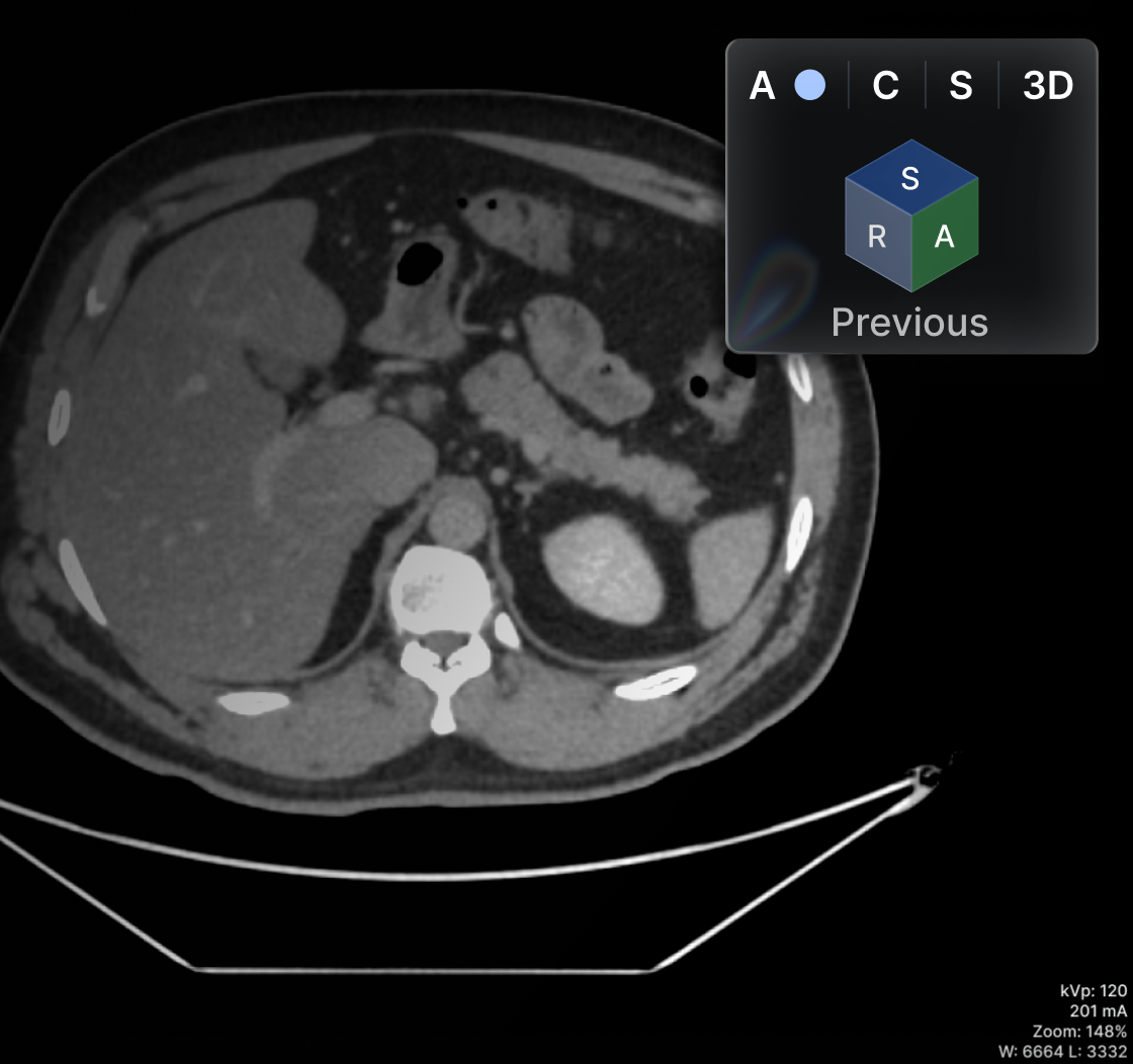
Oblique MPR in the main viewer (not only on the MPR viewer)

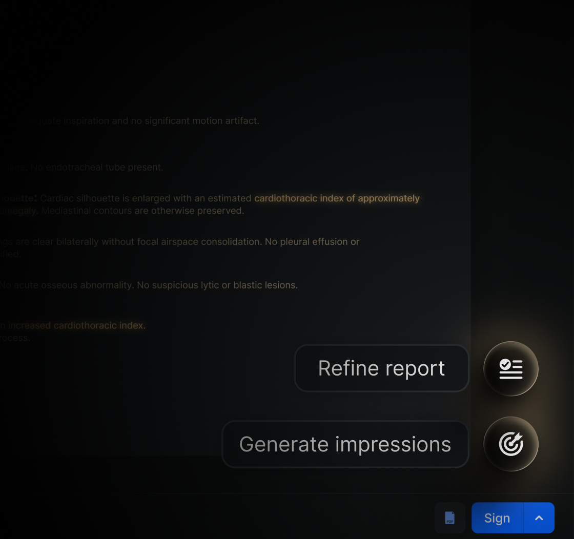
.png)
Browse all
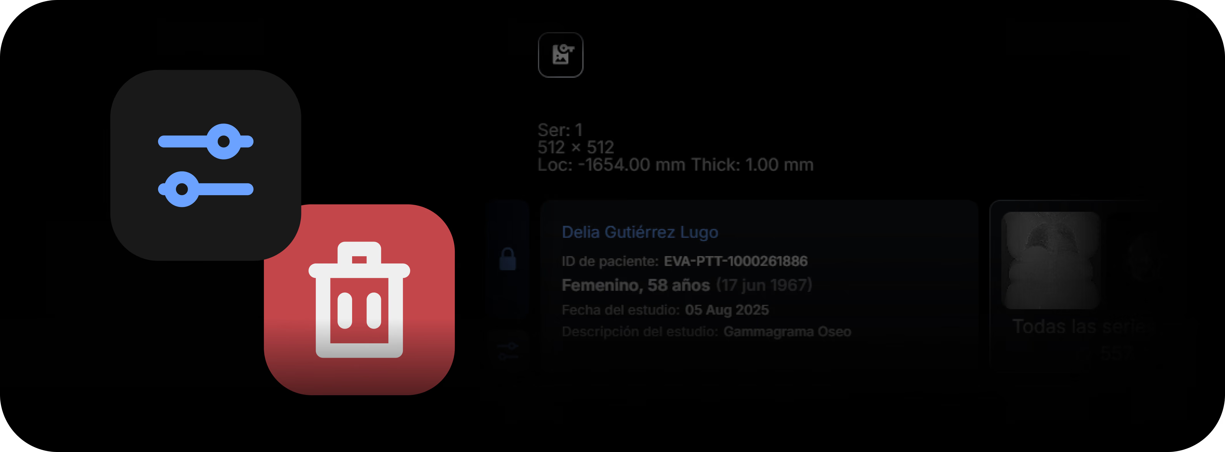
Instantly Eliminate Redundancies with Series and Image Deletion
The platform now incorporates a tool that allows individual images or entire series to be deleted directly from the PACS viewer, without the need to access external administrative tools or the backend of the system. This functionality is enabled exclusively under permission controls and policies defined by each institution.
What it is
This is a feature available within the viewer that allows authorized users to delete specific images or entire series within a study, in a secure and audited manner. Deletion applies to duplicate images, corrupted series, or content that must be removed due to acquisition errors or regulatory reasons.
What it's for
It solves common operational scenarios such as:
- Deleting duplicate series sent by mistake.
- Removing incomplete, corrupted, or poorly reconstructed images.
- Correcting erroneous submissions before diagnostic interpretation.
- Maintaining DICOM file consistency according to institutional quality policies.
- Complying with deletion requests for privacy or regulatory compliance reasons.
How it affects the workflow
Previously, deleting images or series required intervention from the PACS administrator or technical support through the RIS, causing delays and operational tickets. Now, clinical teams can manage simple corrections directly from the viewer without slowing down the reading workflow or affecting the diagnostic schedule. All actions are logged in the audit trail with full traceability.
What changes compared to previous versions
Previously, there was no way to delete DICOM content from the viewer. This process was only possible through administrative modules or scripts on the PACS server. With this update:
- Deletion is enabled directly from the viewer.
- Visible controls are provided per series or image.
- Mandatory confirmation is required before deleting.
- The entire process is audited and reversible according to the client's retention policy.
Implications for the client
The use of this tool is subject to each institution's internal medical data governance policies. This module is activated only by roles and permissions, defined in coordination with the client's system administrator. It is not enabled by default for all users.
Final considerations
- Deletion is a critical action and requires users with elevated permissions.
- Every deletion is logged with user, date, time, study, and reason.
- It is recommended to enable this tool only for users responsible for quality control or PACS management.
- The feature complies with traceability regulations, DICOM standards, and best practices in medical image management.
- It can be integrated with retention and recovery policies according to the client's configuration.
Have you tried it yet? If you have any questions, our team of experts is ready to help you in the Eden PACS support chat, or by email at soporte@edenmed.com
.png)
Collaborate Frictionlessly with the Interpretation-in-Progress Alert
As part of the collaborative control tools in the reading workflow, the RIS-PACS system incorporates the Interpretation in Progress feature, designed to prevent conflicts when multiple users attempt to report on the same study at the same time. This feature ensures report integrity, eliminates duplicate work, and maintains traceability of who has a study in use.
What it is
It is an automatic locking mechanism that activates the moment a radiologist opens a study to generate a report. While the reporting session is active, the system identifies the user as the temporary owner of the interpretation and restricts other users' access to the same reporting workflow.
If another user attempts to open the study, the system displays a notice indicating that the report is in progress and shows the name of the radiologist who is interpreting it.
What it's for
The interpretation-in-progress feature allows you to:
- Prevent two radiologists from generating simultaneous reports on the same study.
- Maintain clear traceability of who is working on a case in real time.
- Reduce the risk of information loss or contradictory versions in reports.
- Facilitate coordination in distributed work teams or overlapping shifts.
How it affects the workflow
Previously, it was possible for more than one user to open the same study and start a report in parallel, generating duplications and consistency errors. With this feature, the system prevents overlap by locking editing for other users while the interpretation is active.
Other radiologists can view the study in read-only mode, but will not be able to modify it or generate a report until the lock is released.
What changes compared to previous versions
- An automatic editing lock is activated when a user opens a study to report on it.
- Other users receive a notice that the report is already in progress, along with the name of the assigned radiologist.
- The study remains available in read-only mode for anyone who consults it, but is not editable.
- The lock is released as soon as the radiologist closes the report or ends the session.
Implications for the user
The radiologist who opens the study first acquires temporary ownership of the interpretation, ensuring that only one report is under construction at a time. Other users can monitor the study's status, but must wait until it is released to begin their own participation, if applicable. The institution gains greater security and traceability in the management of clinical reports.
Final considerations
The interpretation-in-progress feature reinforces system reliability in collaborative, high-volume environments. Its use eliminates frequent operational errors and ensures that each report is linked to a clear owner.
Have you tried it yet? If you have any questions, our team of experts is ready to help you through the Eden PACS support chat, or by email at soporte@edenmed.com
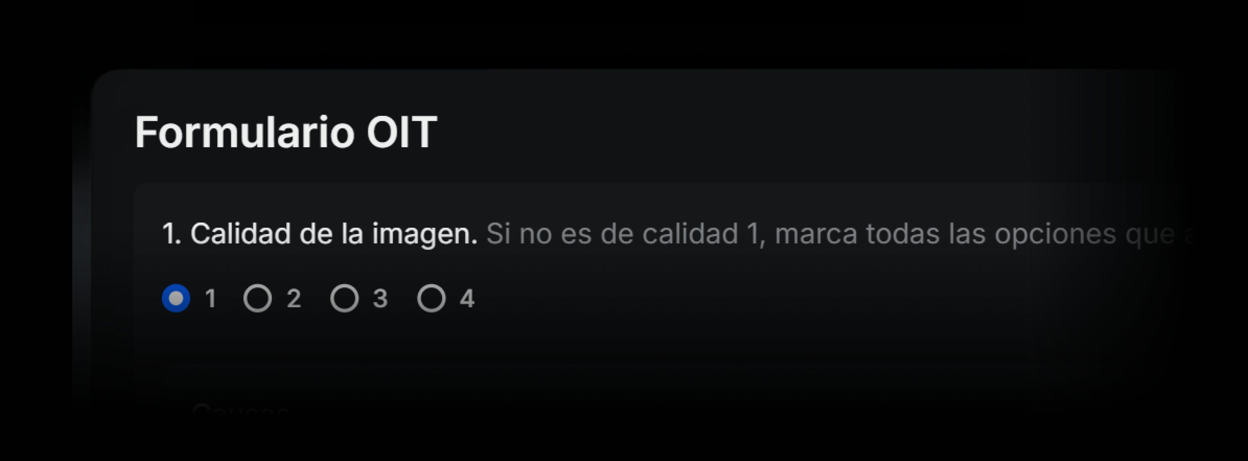
Interpret Under International Standards with the ILO Form
ILO form for pneumoconiosis classification
As part of the strengthening of structured reading and specialized protocols, the ILO (International Labour Organization) Classification of Radiographs of Pneumoconioses form has been incorporated directly into the reading workflow of Eden RIS-PACS. This tool enables standardized, comprehensive capture of radiographic findings associated with occupational exposure to inorganic dusts, following the international guidelines of the ILO/ICRP system.
What it is
This is a structured form that opens within the reporting panel and allows the radiologist to record all fields required by the ILO system: profusion of small opacities, shape and size, large opacities, pleural abnormalities, other findings, and additional notes. It includes guided selection through drop-down lists, standardized codes, and consistency validation.
The tool allows the reader's identification and the study identification to be entered, and facilitates completion for both diagnostic and occupational screening studies. Results can be exported as part of the radiology report or as a structured supplementary file for epidemiology or quality control.
⚠️ Interpretation of radiographs using this form must be performed exclusively by radiologists trained and certified in ILO reading, through courses endorsed by the International Labour Organization.
What it is for
The form is especially useful for:
- Studies requested by labor entities, insurers, or public health institutions.
- Population screening in mining, construction, or asbestos-exposed areas.
- Clinical audits and quality control in pneumoconiosis readings.
- Standardized longitudinal capture of pulmonary radiographic findings associated with chronic exposure.
How it affects the workflow
Previously, radiologists had to rely on external PDF forms or write non-standardized free-text descriptions. With the new tool, completion is done directly from the viewer, in parallel with the narrative report, reducing errors, data entry time, and loss of critical information.
The data is structured, auditable, and exportable, allowing its use in epidemiological databases, external validation, or integration with occupational modules.
Implications for the user
The form is optimized for fast reading through drop-down lists and automatic validations. It can be integrated as a standard template in studies with an occupational protocol. Its use is recommended exclusively in studies with suspected or monitored chronic dust exposure, especially pneumoconiosis caused by silica, coal, or asbestos.
The interface is designed to ensure that no required field is omitted, and it allows final comments to be added for additional clinical observations not covered by the classification.
Final considerations
The incorporation of the ILO form within Eden raises the standard of occupational reading, improves traceability of suspected pneumoconiosis cases, and facilitates compliance with international protocols in high-risk environments. It is a key tool for institutions with a significant volume of occupational reading or an interest in occupational public health.
Have you tried it yet? If you have any questions, our team of experts is ready to help you in the Eden PACS support chat, or by email at soporte@edenmed.com
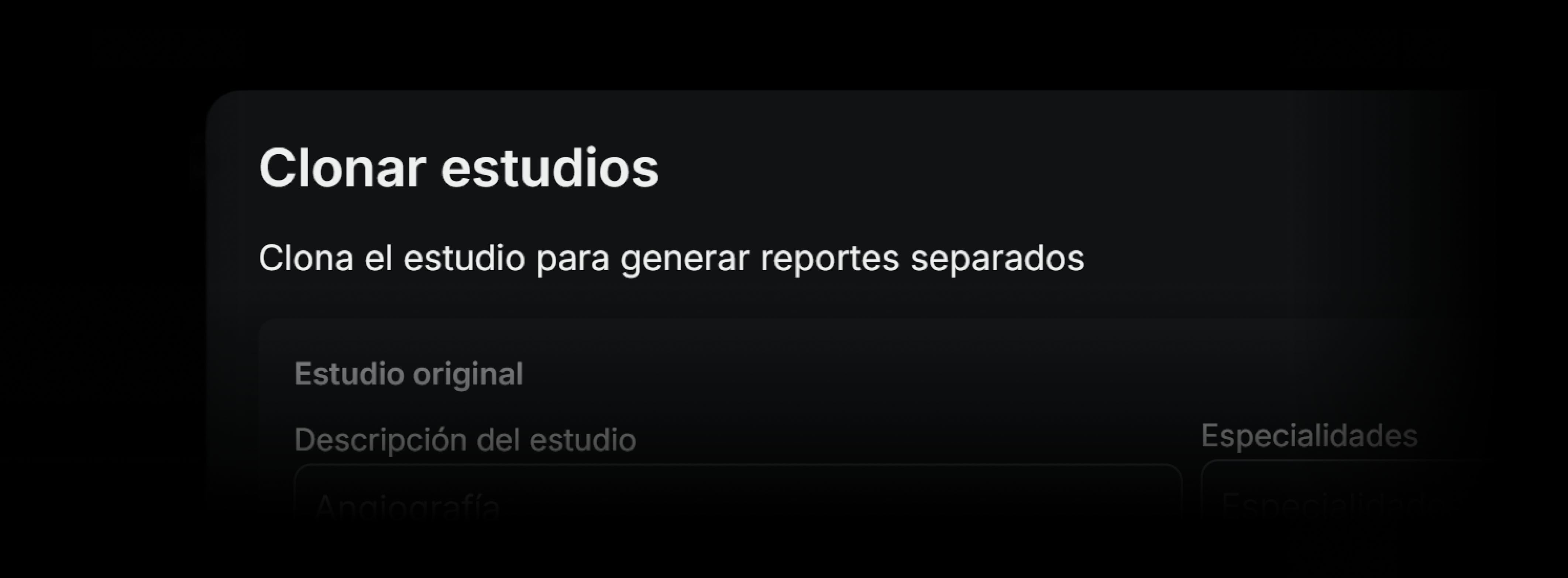
Deliver Specialized Results with Study Cloning
As part of the enhancements to the RIS-PACS system, the ability to clone a study has been added, allowing the generation of independent copies, each with its own report. This functionality provides greater clinical flexibility when reading studies that cover multiple anatomical regions or need to be addressed by different subspecialties.
What it is
This is a tool that allows the radiologist to duplicate a study from the reporting panel. Each generated clone behaves as an independent study: it can have its own study type, specialty, report status, and signature, without altering the original. The system allows the same study to be cloned multiple times if needed.
During the process, the user can assign a new name to each clone, select the corresponding specialty, and decide whether to keep or delete the original report. If the study does not yet have a report started, the option to keep the original report appears disabled.
When a study, due to operational design, was not separated at the time of acquisition or through the sending workflow from the technologist, it is now possible to do so directly from the viewer, without the need for reprocessing in PACS or resending from the technical console.
What it is for
The cloning tool is especially useful for:
- Radiologists who receive whole-body or multi-region studies and wish to report them separately.
- Subspecialists who wish to divide studies by organ or system (for example, one clone for the chest and another for the abdomen).
- Cases where multiple radiologists need to sign different sections of the same original study.
- Preparing independent versions for research, teaching, or external review.
How it affects the clinical workflow
Previously, radiologists had to report long or complex studies in a single report, making organized reading by region difficult. Now, with cloning, studies can be separated according to clinical logic without depending on the technical origin. This improves report traceability, subspecialty assignment, and clarity for the referring physician.
The original study remains as a reference, but each clone follows its own path for signature, status, and interpretation. It can also have its own images, key, and findings.
What changes compared to previous versions
Basically, this possibility did not exist before, and users needed to carry out the process from the RIS through the administrator.
Implications for the user
The cloning feature changes the way radiologists can organize and distribute the reading of complex studies. Instead of forcing a single report with multiple findings, it is now possible to separate content in an orderly way, facilitating specialization, distributed signing, and clinical prioritization.
It also eliminates the need for technical reprocessing when a study was not correctly segmented from the console. This represents an operational saving and an improvement in the clinical user experience.
Final considerations
The cloning tool is designed to improve the organization and traceability of the medical report, especially in scenarios of high anatomical complexity or collaborative work. Its implementation should be accompanied by clear protocols for naming and assigning clones, so that the integrity of the original study is not lost.
The correct use of cloning can result in higher report quality, better distribution of work among subspecialists, and greater clarity for the treating physicians.
Have you tried it yet? If you have any questions, our team of experts is ready to help you in the Eden PACS support chat, or by email at soporte@edenmed.com
.png)
Speed Up Clinical Comparisons with Anatomical Synchronization
As part of the advanced navigation tools in the medical imaging viewer, the RIS-PACS system incorporates the anatomical synchronization feature. This allows the radiologist to simultaneously navigate to the same anatomical region across different series, modalities, or studies, facilitating comparison and improving efficiency in interpretation.
What It Is
Anatomical synchronization is a viewer capability that detects the anatomical correspondence between two or more series or studies and automatically adjusts the viewpoint, slice, and orientation of all linked images, regardless of differences in acquisition, slice thickness, or protocol.
It can be applied to comparisons within the same study (e.g., T1 and T2 in MRI) or between different studies (e.g., prior CT vs. current), allowing a pan or zoom in one series to be replicated at the equivalent location in the others.
What It's For
Anatomical synchronization enables:
- Directly comparing findings between studies with different acquisitions.
- Reducing the time spent manually searching for the same region in prior studies.
- Improving accuracy in evaluating subtle changes in lesions or structures.
- Ensuring anatomical alignment, even with different patient positions or technical parameters.
- Integrating comparison with other tools such as synchronized scroll, zoom and WWWC
How It Affects the Workflow
Previously, the radiologist had to manually navigate each series or study to find the same anatomical level or region, which generated more clicks, movements, and risk of error in comparisons. Now, with anatomical synchronization, a single movement in one series moves all the others to the equivalent point, maintaining orientation and scale, and avoiding loss of reference. This speeds up the review of comparative studies, especially in contexts of oncological follow-up, postsurgical monitoring, or progression of chronic diseases.
Implications for the User
The radiologist can enable or disable anatomical synchronization as needed. The feature is compatible with other viewer synchronizations (scroll, zoom, WWWC) and can be configured to work only between specific series or between complete studies.
For optimal performance, studies must have complete and consistent DICOM data in the position, orientation, and spacing fields, or spatial registration maps when different modalities are used.
Final Considerations
Anatomical synchronization is a key tool for environments that require fast, precise comparative reviews. It does not replace clinical judgment, but it improves the speed and consistency of reading, especially in reviews involving multiple studies and modalities. It is activated instantly from the viewer's control panel and requires no additional software installation.
Have you tried it yet? If you have any questions, our team of experts is ready to help you in the Eden PACS support chat, or by email at soporte@edenmed.com
.avif)
Build Your Ideal Workspace: Customize Your Columns in Eden PACS
As part of the worklist module update, the RIS-PACS system now offers the ability to customize the display of each list through a simple menu that allows columns to be shown, hidden, and dynamically reordered. This feature aims to improve the user experience and adapt to the preferences of each radiologist or operations coordinator.
What It Is
It is a tool built into the worklist configuration menu that offers two main capabilities:
- Selecting which columns are shown and which are hidden, based on the relevance of each role.
- Reordering the columns by dragging them within the same menu, without needing to leave the work screen or resort to advanced settings.
Applied changes take effect immediately and are saved as a user preference, ensuring that the list is always displayed with the same customized layout.
What It's For
Column reordering allows each user to adjust the list interface according to their priorities. It is especially useful for:
- Radiologists who want to see key clinical information first (e.g., priority, institution, modality).
- Coordinators who need to highlight SLA, assignment, or operational status.
- Multisite teams that handle large volumes of studies and require clearer on-screen filters.
- Fast-reading contexts where minimizing navigation steps speeds up interpretation.
How It Affects the Workflow
Previously, the study list had a fixed column structure that did not always meet each user's needs. Now, with this new tool, each radiologist can organize the view according to their own workflow logic, which reduces search time, facilitates study prioritization, and improves individual productivity.
What Changes Compared to Previous Versions
Previously, there was no way to customize the columns directly from the viewer. Changes had to be requested from an administrator or were limited to global settings. With this new feature, the end user has full control and can reorganize their interface immediately and independently.
Implications for the User
The user gains greater control over their work environment, adapting the interface to their specific needs without relying on technical support. This allows each radiologist to work with their own layout, while the institution maintains full traceability of studies.
Final Considerations
Column reordering turns the worklist into a flexible, customized tool that adapts to both individual radiologists and coordinators of large teams. Its use facilitates clinical prioritization, improves visibility of critical data, and optimizes efficiency in study management.
Have you tried it yet? If you have any questions, our team of experts is ready to help you via Eden PACS support chat, or by email at soporte@edenmed.com
.avif)
Drive Institutional Excellence with Peer Learning
As part of Eden PACS's clinical excellence mechanisms, a new layer of control has arrived that allows peer review of studies to ensure diagnostic quality and continuous learning.
This feature ensures clear traceability of physician performance and institutional standards, and serves as an unprecedented competitive advantage for institutions of any size.
What it is
The peer learning tool enables peer reviews to be activated for any study within the institution. The assigned physician can access the study to fill out a digital form tailored to the institution's evaluation needs. Afterward, the user who originally interpreted the study will receive the feedback by email.
What it's for
- Ensuring clinical quality in the diagnoses generated by your institution.
- Maintaining traceability and compliance with diagnostic standards.
- Fostering the exchange of knowledge among colleagues.
- Generating metrics to evaluate clinical or contractual performance.
Workflow
Previously, peer feedback only took place in academic settings, with manual, exhaustive forms that disrupted the workflow. Now, with peer learning, any institution can implement digital feedback systems to ensure the highest quality standard.
Users receive feedback directly by email. The result of each review is automatically logged and can be viewed in operational reports or dashboards focused on quality.
Implications for the user
By having access to peer reviews without significant adjustments to their workflow, radiologists can ensure diagnostic quality and continue the learning process expected of their profession. Institutions can use these results to validate clinical performance, ensure excellence and increase their study volume.
Peer learning is a key tool for improving the quality of radiological service, especially in settings with multiple radiologists, differentiated contracts, or demanding institutions where excellence is a competitive advantage.
Its implementation does not require changes to the viewer or the reading console. It can be configured from the RIS/PACS administration panel and adjusted according to the institution's needs.
Have you tried it yet? If you have any questions, our team of experts is ready to help you in the Eden PACS support chat, or by email at soporte@edenmed.com
.png)
Minimize Your Film Consumption with On-Demand Printing
Eden PACS has incorporated a new on-demand printing tool that allows images to be exported and printed directly from the reading environment, without needing to leave the viewer or use intermediate stations. This feature is designed to provide greater autonomy, precision, and flexibility when documenting or sharing findings.
What it is
It is a tool integrated into the viewer that allows the user to generate customized printouts of DICOM images in different formats, sizes, and resolutions. From the top menu, users can access the printing options, select the orientation and paper type, and export directly without loss of quality.
This way, the process is under the user's full control, reducing the use of duplicate films and minimizing printing to only when the physician or patient requires it.
What it's for
On-demand printing allows you to:
- Document findings for clinical reference or a second opinion.
- Include selected images in patient reports.
- Prepare deliverables for patients who require them.
- Support material for academic presentations or interdisciplinary sessions.
In addition, this feature is especially useful in modalities where printed images are still used as visual support, such as mammography, interventional radiology, orthopedics, and oncology.
How it affects the workflow
Previously, printing an image required exporting it manually, opening other software, or relying on specific stations. Now, everything can be done from the viewer, without interruptions or loss of quality.
This speeds up the workflow, reduces unnecessary steps, and gives the user greater control over the final format. It also allows for a faster response to requests from patients, referring physicians, or administrative situations.
What changes compared to previous versions
- Printing no longer depends on external stations or intermediate exporting.
- The user can choose standardized sizes (Letter, A4, DICOM 14x17, among others) and adjustable resolutions.
- Alignment options, label display, and compression levels are included.
- The process is immediate and integrates into the reading workflow without affecting viewer performance.
Implications for the user
Users gain autonomy in preparing visual material, without relying on technical support or additional steps. The feature is available for all studies, provided export or printing permissions are enabled.
It is advisable to define internal policies on what type of printouts are allowed, with what level of identification, and for what clinical or administrative purposes.
Final considerations
On-demand printing is a tool designed to meet the real needs of the modern clinical environment: fast, configurable, and user-centered. It allows images to be exported with diagnostic quality, in formats compatible with paper, medical documents, or physical deliverables.
Have you tried it yet? If you have any questions, our team of experts is ready to help you through the Eden PACS support chat, or by email at soporte@edenmed.com
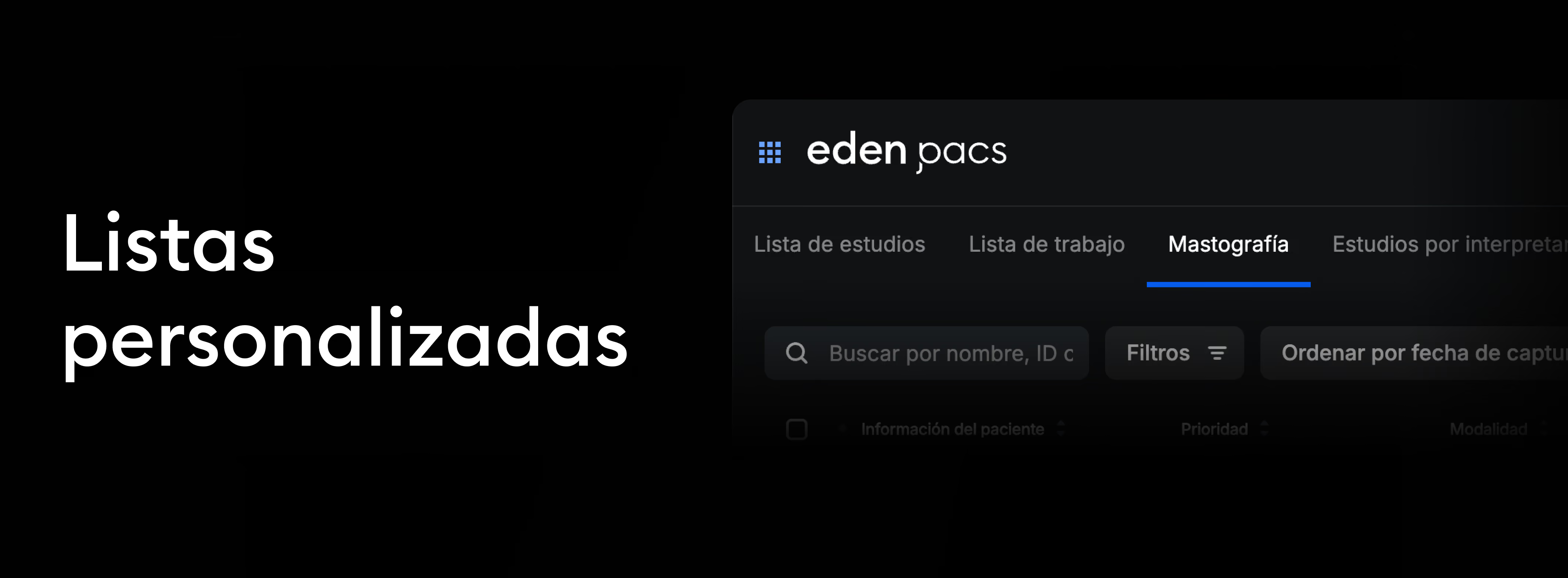
Your Studies, Organized Your Way with Custom Lists
Now, your study lists adapt to you. With the new custom lists feature in Eden PACS, you can filter groups of studies, save your frequent searches, and access them instantly.
It streamlines the radiology workflow and optimizes collaboration between teams, providing real-time visibility of:
- Assigned studies pending sign-off, for individual radiologists.
- Emergency studies, for shift leads in hospital settings.
- Subspecialty studies, for specialist physicians.
- The status of each study, for institutions following operational agreements.
How it affects the workflow
Previously, users accessed a generic list that required applying manual filters each time. Now, they can have smart, persistent lists that load automatically with a single click. This reduces operational time, improves visibility of study status, and allows for more efficient workload distribution among teams.
Each user can create multiple lists and switch between them according to their needs without losing previously configured filters.
There are two types of custom lists in Eden PACS:
- Institutional lists: configured by the organization for all its users, optimizing workflows and ensuring adherence to standardized protocols. These lists can only be edited or deleted with the support of the Eden technical team.*
- Personal lists: created by the user according to their preferences and can be viewed, modified, or deleted at any time. These lists will only appear for the user who created them, and will not be visible to other users in the organization.
*To request the creation of an institutional list, contact our experts at soporte@edenmed.com
How to create your own custom list
- Navigate to your study list and click the "Filters" button. In the filtering modal, choose your search criteria.
- At the bottom left of the modal, click "Save as custom list".
- Give your list a name and save it. Now, you'll be able to access it with a single click every time you log into your Eden PACS account.
If you want to edit your custom list, simply click "Filters".
- To adjust the list, select the filters you want to display and click "Save changes to list".
- To delete the list, click the "Delete" button.
It facilitates the traceability, prioritization, and coordination of key studies, optimizing radiology operations in highly complex environments. Start creating custom lists today in Eden PACS.
Have you tried it yet? If you have any questions, our team of experts is ready to help you via the Eden PACS support chat, or at soporte@edenmed.com

Interpret Without Delays with Study Preloading in Eden
As part of the viewer optimization and improved radiologist experience, we have implemented a study preloading tool. This feature allows studies to be selected directly from the Worklist for early download to the local server or viewer cache, achieving speeds up to 100 times faster and reducing opening times during reading.
What it is
A feature that enables proactive downloading of one or several studies from the Worklist without needing to open them immediately. This way, when the radiologist accesses the study, the images are already available locally, minimizing load latency, especially over remote connections or with complex studies (MRI, multi-series CT).
What it is for
- Reduces waiting times during reading.
- Optimizes the radiologist's session by preparing upcoming cases.
- Increases productivity on high-volume days.
- Facilitates the reading of heavy modalities, such as MRIs, mammograms, or multiphase studies.
Impact on workflow
Previously, each study was downloaded upon opening, causing delays. Now, the radiologist or technologist can preselect the studies to be read in the next session, ensuring instant availability when needed.
It is especially useful in hybrid environments (cloud + local) or in teleradiology, where download speed directly affects efficiency.
Changes to your PACS
- The Worklist preloads automatically upon logging into Eden PACS.
- New column to preload or remove the preload of all studies simultaneously.
- Option to manually select and preload studies in the Worklist, study lists, or custom lists.
- Visual indicators of download status:
- Gray: Not preloaded.
- Blue: In progress.
- Green: Preloaded.
- The cache is managed automatically, freeing up space after studies are signed off or after a 24-hour period. The download will also be removed if the user logs out of their Eden account.
Implications for the user
Preloading offers a smoother experience, eliminating dead time and allowing the radiologist to focus on interpretation.
Important: Preloading is local to a single device; if you switch workstations, studies will need to be preloaded again. It does not alter study content, only makes it available earlier.
Usage recommendations
- Heavy studies or those with multiple series.
- Case lists for batch reading (e.g., shift planning).
- Scenarios with limited connectivity, where every second impacts productivity.
Have you tried it yet? If you have any questions, our team of experts is ready to help you via the Eden PACS support chat, or at soporte@edenmed.com

Maximize Your Workspace with the New Horizontal Bar
In Eden PACS, you own your space. Now you can choose how to view the new and improved horizontal series bar, designed to streamline your radiological interpretation.
Dynamic bar
The new series bar is located at the bottom of the DICOM viewer and remains hidden by default, creating more display space to view the full image without obstructions.
- To navigate the studies bar, simply move your cursor toward the study tab in the bottom bar.
- If you want to keep the bar visible, click the lock icon to pin it. This way, you can maintain visibility of all images.
Series, all-in-one
The new bar also includes a combined series that brings together all the images in the study so you can view them all at once, in the same place, with just a scroll.
Related studies
The series bar also lets you view all studies generated for the same patient*, making it easier to compare previous studies for a more complete interpretation.
Simply navigate to the study you want to review and click the series you want to see. In the additional actions section (፧) you can:
- View and/or edit the study information
- Open the DICOM study in a new window
- View additional study files
- Download the study in DICOM format
- Download the study in PDF format
Identification errors
In addition, you will be able to identify studies with "identification errors." This feature detects when the patient information partially matches your current case but shows discrepancies such as phone number or ID. This way, you can correct capture errors without losing access to studies that could be relevant to the diagnosis.
Your interpretation, now more agile, precise and complete, all from Eden PACS. Discover the new series bar today in your DICOM viewer, or request it from your nearest representative.
Have you tried it yet? If you have any questions, our team of experts is ready to help you in the Eden PACS support chat, or by email at soporte@edenmed.com
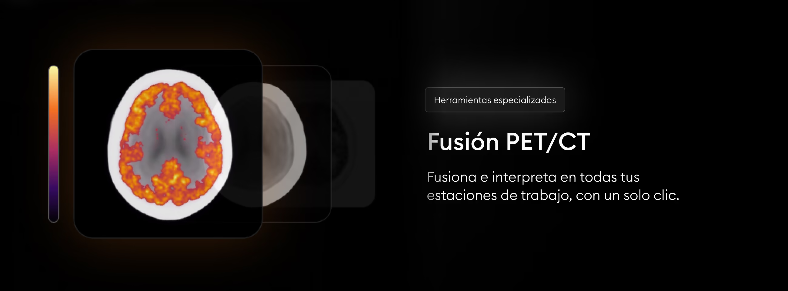
Reveal the Invisible Instantly with PET/CT Fusion in Eden PACS
Make the invisible visible with a single click. Use the PET/CT Fusion tool to combine the metabolic information of a PET with the anatomical precision of a CT, visualizing critical details for the highest standard of diagnosis.
Metabolic and anatomical information in one view
Available on all your workstations
Use PET/CT Fusion instantly across all your workstations, at no cost for additional licenses or local equipment. All from Eden PACS.
No papers match your search.